A net for everyone fully personalized and unsupervised neural networks trained with longitudinal data from a single patient
2025-04-30
0
0
536.54KB
15 页
10玖币
侵权投诉

“A net for everyone”: fully personalized and unsupervised
neural networks trained with longitudinal data from a single
patient
Christian Strack1,2*, Kelsey L. Pomykala3, Heinz-Peter Schlemmer1,5, Jan Egger3,4
Jens Kleesiek3,4,5
1 Division of Radiology, German Cancer Research Center (DKFZ), 69120 Heidelberg, Germany
2 Medical Faculty Heidelberg, Heidelberg University, 69120 Heidelberg, Germany
3 Institute for AI in Medicine (IKIM), University Hospital Essen (AöR), Girardetstraße 2, 45131
Essen, Germany
4 Cancer Research Center Cologne Essen (CCCE), University Medicine Essen, Hufelandstraße
55, 45147 Essen, Germany
5 German Cancer Consortium (DKTK), Partner Site Essen, Hufelandstraße 55, 45147 Essen,
Germany
*corresponding author: c.strack@dkfz-heidelberg.de
Abstract
With the rise in importance of personalized medicine, we trained personalized neural networks to
detect tumor progression in longitudinal datasets. The model was evaluated on two datasets with
a total of 64 scans from 32 patients diagnosed with glioblastoma multiforme (GBM). Contrast-
enhanced T1w sequences of brain magnetic resonance imaging (MRI) images were used in this
study. For each patient, we trained their own neural network using just two images from different
timepoints. Our approach uses a Wasserstein-GAN (generative adversarial network), an
unsupervised network architecture, to map the differences between the two images. Using this
map, the change in tumor volume can be evaluated. Due to the combination of data augmentation
and the network architecture, co-registration of the two images is not needed. Furthermore, we
do not rely on any additional training data, (manual) annotations or pre-training neural networks.
The model received an AUC-score of 0.87 for tumor change. We also introduced a modified
RANO criteria, for which an accuracy of 66% can be achieved. We show that using data from just
one patient can be used to train deep neural networks to monitor tumor change.
Keywords: Neural networks, personalized, Wasserstein-GAN, unsupervised, machine learning,
privacy-safe, zero-training data, longitudinal, brain tumor, MRI.
1 Introduction
One key difference between human and artificial intelligence is the number of training examples
needed to generate knowledge. While children can learn to recognize new objects with only a few
examples, most machine learning tasks require hundreds of examples for the same task. In fact,
increasing the dataset size is often a key step in improving the performance of a machine learning
model. ImageNet [1], the most famous dataset in computer vision, now consists of over 14 million
training examples. The state of the art models in computer vision are often trained on large
datasets such as ImageNet and may not transfer well to smaller datasets. Getting large datasets
may not always be a feasible approach though, especially in the medical domain.
Gathering large datasets is one of the key challenges of medical deep learning applications.
Keeping a patient’s medical information safe is critical and there are laws protecting it in most
countries. This makes it more difficult to get the data and leads to the medical datasets being
much smaller compared to traditional computer vision datasets. Additionally, deep neural
networks themselves offer another privacy threat. It has been shown that training examples of
fully trained networks can be recovered with a model inversion attack [2]. This makes it more
difficult to publish medical deep learning applications as the patient’s privacy can not be
guaranteed. These two reasons give a big incentive to find ways to train neural networks with
smaller datasets or even just one patient’s data.
There have been several models proposed to challenge the task of reducing the number of
training examples. One shot learning is a method of learning a class from only one labeled
example [3]. Siamese neural networks are able to determine if two images show the same person,
even if they have never seen images of that person before [4]. They have also been used in
medicine to distinguish between COPD and asthma [5]. While new classes can be learned from
as little as one example, one shot learning still requires thousands of training examples of other
classes beforehand. Another method to handle small datasets is transfer learning, where
networks trained on large datasets are used as a starting point to train on training examples of
new classes. Transfer learning makes use of the fact that features learned on the large dataset
can be reapplied to new data.
In this paper, we introduce personalized neural networks, which use only one patient’s data for
training. Our proposed method only needs two MRIs from the same patient and no additional
pretraining. This also results in a privacy-safe processing of the data, because the data “stays”
within the same patient. Our model is based on Generative Adversarial Networks (GANs) [6].
GANs have gained in popularity in recent years in the medical AI community. Originally used for
image synthesis, there have been applications to generate medical images [7, 8]. Other studies
focus on classification or segmentation tasks [9, 10]. We apply the personalized neural networks
on subjects with brain tumors.
Brain tumors belong to the most devastating diagnoses, in particular for a confirmed glioblastoma
multiforme (GBM) [11]. Despite massive research efforts and advancements in other cancer
types, like breast cancer [12] or prostate cancer [13], the life expectancy of a confirmed GBM with
treatment, including chemotherapy, radiotherapy and surgery, is still only around one year [14].
Nevertheless, disease progression and treatment decisions are strongly dependent on maximum
tumor diameter and tumor volume, as well as the corresponding morphological changes during a
treatment period. The imaging method of choice here is magnetic resonance imaging (MRI).
However, MRI does not provide any semantic information for brain structures or the brain tumor
per se. This has to be done manually, semi-manually or automatically, in a post-processing step,
commonly referred to as a segmentation. Manually performed, however, a segmentation is very
time-consuming and operator-dependent, especially when performed in a three-dimensional
image volume [15], which needs slice-by-slice contouring. Hence, an automatic (algorithmic)
segmentation is desired, especially when large quantities of data volumes have to be processed.
Even if it is still considered an unsolved problem, there has been steady progress from year to
year; and data-driven approaches, like deep neural networks, currently provide the best (fully
automatic) results. However, a segmentation with a data-driven approach, like deep learning [16],
comes with several burdens: Firstly, the algorithm generally needs massive annotated training
data. Additionally, for inter-patient disease monitoring, several segmentations have to be
performed, and in addition, these scans have to be registered to each other (which also adds
uncertainty to the overall procedure, especially when deformed soft-tissue comes into play [17]).
In this regard, we want to tackle these problems with a personalized neural network that needs
just the patient’s data, no annotations and no extra registration step. To the best of our knowledge,
this is the first study using this little training data to train a deep neural network in the medical
domain. The method addresses the issues of gathering big datasets in medicine and producing
a privacy-safe network. The approach is considered as unsupervised learning as no data
annotation is necessary. We evaluate the model with an ROC analysis as well as modified RANO
criteria on two different datasets of longitudinal MRI images of patients with glioblastoma.
2 Methods
2.1 Model architecture and training
The neural network architecture used in this study is based upon Wasserstein GANs [18]. This is
a modified version of Generative Adversarial Networks (GAN) [6]. These are a form of deep neural
networks in which two sub-models are trained adversarily in a sum-zero game. A generator is
trained to create new images, while a discriminator is trained to distinguish between real and
synthetic images. In Wasserstein GANs the discriminator is modified to a critic function which
leads to more stable training [18].
Our network architecture is similar to the model used by Baumgartner et al [19]. The aim of the
network is to create a map which transforms an image from the first timepoint (t1) to the second
timepoint (t2). This will make the model learn to represent the changes between the images, more
specifically tumor growth/reduction in our case. To do this, augmented versions of the image at
t1 are used as input to the generator. The generator will try to create a map that, when added to
the input image creates an image of t2. The critic will try to distinguish these generated synthetic
t2 images from the real t2 images. Thereby forcing the generator to learn the differences between
the two timepoints.

摘要:
展开>>
收起<<
“Anetforeveryone”:fullypersonalizedandunsupervisedneuralnetworkstrainedwithlongitudinaldatafromasinglepatientChristianStrack1,2*,KelseyL.Pomykala3,Heinz-PeterSchlemmer1,5,JanEgger3,4JensKleesiek3,4,51DivisionofRadiology,GermanCancerResearchCenter(DKFZ),69120Heidelberg,Germany2MedicalFacultyHeidelber...
声明:本站为文档C2C交易模式,即用户上传的文档直接被用户下载,本站只是中间服务平台,本站所有文档下载所得的收益归上传人(含作者)所有。玖贝云文库仅提供信息存储空间,仅对用户上传内容的表现方式做保护处理,对上载内容本身不做任何修改或编辑。若文档所含内容侵犯了您的版权或隐私,请立即通知玖贝云文库,我们立即给予删除!
相关推荐
-
【词汇变形总汇】2025高考词汇变形总汇 - 教师版VIP免费
 2024-12-06 8
 -
【超简37页】新课标高考英语考纲3500词汇VIP免费
 2024-12-06 18
 -
《高考英语3500词详解》(WORD版)VIP免费
 2024-12-06 33
 -
《高考英语3500词详解》VIP免费
 2024-12-06 29
 -
高中英语-[教师版]80天通关高考3500词汇VIP免费
 2024-12-06 34
 -
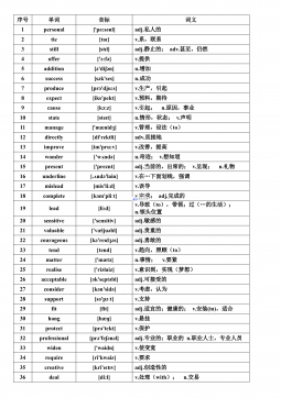
高中人教选修7课文逐句翻译VIP免费
 2024-12-06 15
 -
高中人教选修7课文原文及翻译VIP免费
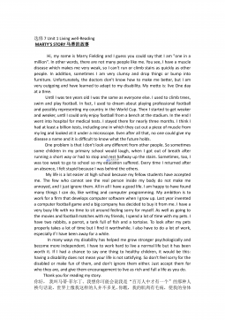 2024-12-06 41
 -
高中人教必修4课文逐句翻译VIP免费
 2024-12-06 40
 -
高中人教必修4课文原文及翻译VIP免费
 2024-12-06 60
 -
高考英语核心高频688词汇VIP免费
 2024-12-06 34
分类:图书资源
价格:10玖币
属性:15 页
大小:536.54KB
格式:PDF
时间:2025-04-30
作者详情
相关内容
-
山东省泰安市2025届高三四模检测历史答案
分类:中学教育
时间:2026-03-20
标签:无
格式:PDF
价格:10 玖币
-
山东省临沂市普通高中学业水平等级考试模拟试题地理答案
分类:中学教育
时间:2026-03-20
标签:无
格式:PDF
价格:10 玖币
-
山东省临沂市2025届高三上学期教学质量检测考试暨期中考试(九五联考)数学答案
分类:中学教育
时间:2026-03-20
标签:无
格式:PDF
价格:10 玖币
-
山东省九五高中协作体2025高三年级质量检测(九五联考)语文答案
分类:中学教育
时间:2026-03-20
标签:无
格式:PDF
价格:10 玖币
-
山东省九五高中协作体2025高三年级质量检测(九五联考)数学
分类:中学教育
时间:2026-03-20
标签:无
格式:PDF
价格:10 玖币