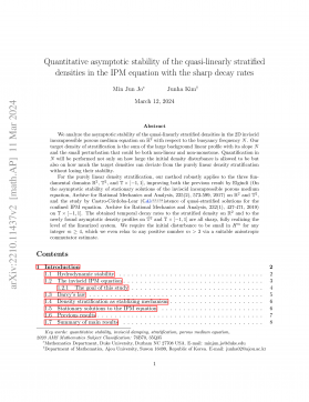
Raman spectroscopic characterization of crater walls formed upon single -shot high -energy femtosecond laser irradiation of dimethacrylate polymer doped with plasmonic gold nanorods

2025-04-29 0 0 518.93KB 17 页 10玖币
侵权投诉
Raman spectroscopic characterization of crater walls formed upon single-shot high-energy
femtosecond laser irradiation of dimethacrylate polymer doped with plasmonic gold
nanorods
István Rigó1, Judit Kámán1,Ágnes Nagyné Szokol1, Roman Holomb1, Attila Bonyár2, Melinda Szalóki3,
Alexandra Borók1,2, Shereen Zangana2, Péter Rácz1, Márk Aladi1, Miklós Ákos Kedves1, Gábor Galbács5,
László P. Csernai1,6,7, Tamás S. Biró1, Norbert Kroó1,8, Miklós Veres1, NAPLIFE Collaboration
1Wigner Research Centre for Physics, Budapest, Hungary
2Department of Electronics Technology, Faculty of Electrical Engineering and Informatics, Budapest University for
Economics and Informatics, Budapest, H1111, Hungary
3Department of Biomaterials and Prosthetic Dentistry, Faculty of Dentistry, University of Debrecen, Debrecen,
Hungary
4Centre for Energy Research, Institute of Technical Physics and Materials Science (MFA), H1121 Budapest, Hungary
5Department of Inorganic and Analytical Chemistry, University of Szeged, Szeged, H-6720 Hungary
6Department of Physics and Technology, University of Bergen, 5007 Bergen, Norway
7Frankfurt Institute for Advanced Studies,Frankfurt/Main, Germany
8Hungarian Academy of Sciences, 1051 Budapest, Hungary
Abstract
The bonding configuration of the crater walls formed in urethane dimethacrylate-based polymer
doped with plasmonic gold nanorods upon irradiation with a single-shot high-energy femtosecond
laser pulse has been studied by Raman spectroscopy. New Raman bands were detected in the 2000-
2500 cm-1 region of the Raman spectrum the intensities of which showed strong dependence on
the concentration of the plasmonic nanoparticles and the energy of the laser pulse. Based on model
calculations of the Raman frequencies of the polymer these peaks were attributed to carbon-
deuterium and nitrogen-deuterium vibrations. Their appearance might indicate the occurrence of
nuclear reactions in the polymer excited by the ultra-strong laser field amplified by the plasmonic
nanoparticles.
Keywords: plasmonic enhancement, femtosecond laser, nuclear reactions, Raman spectroscopy,
dimethacrylate polymer
1. Introduction
The use of high-energy short laser pulses is gaining significance in numerous applications.
Localized surface plasmon polaritons (LSPP) can, for example, be excited efficiently with these
pulses even up to very high laser intensities. One of the reasons to do so is that with the help of
plasmonic nanoparticles the electromagnetic field of the lasers can be amplified on the nanoscale
resulting in fields orders of magnitude higher, than those of the laser pulse. Our motivation to
explore this enhancement effect has been to use these high fields to realize tabletop plasmonic
nano-fusion processes [1]. Ti:Sa femtosecond laser pulses have been used to excite LSPPs in a
polymer sheet, containing resonant plasmonic gold nanorods, with laser intensities up to a few
times 1017 W/cm2. The first step in our studies has been the analysis of the craters formed by
individual laser shots in the material [2]. It was found, that the volume of these craters is always
significantly higher in the polymer containing the gold nanoparticles than in the same polymer
without them. The volume increase can be attributed only to nuclear processes, and it has been
found, that the crater volumes depend linearly on the energy of the laser pulses [2]. Since this
energy production has been found to be comparable, or even larger, than that of the energy of the
laser pulse, it has been decided to use vibrational spectroscopic method (Raman spectroscopy) to
characterize structural changes occurred in the polymeric structure at the crater walls formed upon
irradiation of the material with single-shot high-energy femtosecond laser pulse. The irradiation
with different pulse energies was performed on both a pure structure and also samples doped with
plasmonic gold nanorods (in two different concentrations) having plasmon resonance at the
wavelength of the femtosecond laser, and the Raman measurements were performed on both types
of samples.
Being sensitive to bonding configuration, Raman spectroscopy is widely used to characterize
different polymeric materials, including dimethacrylates. This method can be used to determine
the degree of conversion in these types of polymers, as well, through the changes and ratio of the
Raman peak of the C=C double bonds to that of a reference band belonging to a bond not affected
by the polymerization [3-5]. In addition to the bonds of the polymer frame it allows to study the
different C-H and N-H groups as well. The effect of the laser pulse on the polymer framework,
including the additional conversion upon irradiation in the presence of gold nanoparticles, has been
published earlier [6]. Here we report on the changes observable in the 2000-2600 cm-1 region,
being dependent on the presence of the nanoparticles and the pulse energy as well. In situ laser
induced breakdown spectroscopy (LIBS) measurements were also performed to validate the
Raman findings.
2. Methods
2.1.Materials and sample preparation
The photopolymerizable dimethacrylate resin mixture consists of urethane dimethacrylate
(UDMA) (Sigma Aldrich) and triethylene glycol dimethacrylate (TEGDMA) (Sigma Aldrich) in
3:1 mass ratio. Dodecanethiol-capped gold nanorods (Au-DDT) in size of 25 nm diameter and 75
nm length were purchased from Nanopartz Inc. (part no.: B12-25-700-1DDT-TOL-50-0.25, and
B12-25-750-1DDT-TOL-50-0.25).
The preparation method of pure (UDMA-X) and doped with gold nanorods (UDMA-Au)
polymer samples is described in detail elsewhere [6]. In short, the mixture of the UDMA and
TEGMA monomers and the photoinitiator (and Au-DDT) is placed on a glass slide in a template
allowing to obtain a thin layer of the resin. Then the polymerization mixture is irradiated with a
standard dental curing lamp emitting blue light with 3 min exposure time. The obtained round
shaped thin film samples are clear (UDMA-X non-doped polymer) or have pink color (UDMA-
Au doped samples). The gold nanorods were added to the monomer mixture in 2 different
concentrations of 0.124 m/m% and 0.182 m/m%, and the corresponding samples were marked as
UDMA-Au1 and UDMA-Au2 in this manuscript, respectively. In addition, on some figures, the
laser pulse energy in milliJoules is also added to the sample name, i.e. UDMA-X-25 corresponds
to the crater in the non-doped polymer irradiated with 25 mJ laser pulse.
2.2. Laser irradiation experiments
The irradiation of the samples was implemented by a Ti:Sapphire-based chirped-pulse two-
stage amplifier-laser system (Coherent Hydra) delivering pulses with 40 fs pulse length at 795 nm
central wavelength with 10 Hz repetition rate and 25 mJ maximum pulse energy. The beam was
focused with a lens having 50 cm focal length. The laser irradiation experiments were performed
under vacuum conditions to avoid nonlinear processes in air. The pressure in the vacuum chamber
was in the range of 106 mbar. The sample treatment was achieved by single pulses of different
energy, each illuminating a different region of the sample surface (this was achieved by shifting
the sample laterally after each pulse). Since the energy density is above the ablation threshold of
the material, crater formation was observed at the irradiation spot.
2.3.Raman spectroscopy
A Renishaw InVia micro-Raman spectrometer connected to a LeicaDM2700 microscope
was used for the Raman spectroscopic measurements. The Raman spectra were recorded on the
wall of the formed crater with 532 nm excitation in backscattering geometry, with the laser focused
into a spot of ~2 microns diameter on the sample surface by using a 50X/0.5 NA objective. The
laser power was ~6 mW which equals 5-10% of the maximum intensity of the laser source. The
spectra were recorded in the 1000 - 2650 cm-1 spectral region. Due to the low intensity of the
investigated Raman bands the accumulation time was set to 2 hours in each spot. Before the
measurements, calibration was done by using a silicon wafer and its characteristic peak at 520 cm-
1 Raman shift.
Since this study is about the evaluation and comparison of small-intensity Raman peaks
recorded on different craters formed upon single-shot laser irradiation, a special care was given to
the processing of the measured Raman spectra, that was done with the Origin 2019 software and
a custom Matlab code. The first step was the normalization of the spectra to the C-C peak at 1460
cm-1. Then, background subtraction was performed by using the Asymmetric Least Squares
method [7]. The integral intensity of the Raman peaks was determined by calculating the area
under the curve for the composite Raman band observed in the 2000-2600 cm-1 region.
2.4.Laser induced breakdown spectroscopy
The LIBS measurements were performed in situ, during the single-shot irradiation of the
polymer targets with the high-intensity laser pulses. The laser induced breakdown plasma emission
was collected at 45 degrees angle and collimated by placing a lens at a 4 cm distance from the
target inside the chamber. The collimated beam was then focused into an optical fiber with a core
diameter of 400 μm and transmitted into a high resolution double Echelle spectrometer (having a
4 pm resolution) equipped with an ICCD camera (Demon, LTB, Berlin). The spectrum recording
was done using a fixed gate width of 1 µs and a gate delay of 0.6 μs. The spectrometer was centered
around 656 nm and recorded the spectrum in a 3 nm spectral window.
2.5.Modeling and calculations of the selectively deuterized UDMA monomer
The chemical structure of urethane-dimethacrylate (UDMA) monomer is shown in
Figure 1. Part of this structure containing N-H and C-H2 groups was selected as a starting geometry
for further modeling and subsequent calculations. For better description of the chemical
environment, the dangling chemical bonds at the ends of the model were terminated with methyl
(CH3) and hydroxyl (OH) groups, as shown in Figure 2.
Figure 1. Chemical structure of UDMA monomer together with the part selected for further
modeling and calculations.
摘要:

Ramanspectroscopiccharacterizationofcraterwallsformeduponsingle-shothigh-energyfemtosecondlaserirradiationofdimethacrylatepolymerdopedwithplasmonicgoldnanorodsIstvánRigó1,JuditKámán1,ÁgnesNagynéSzokol1,RomanHolomb1,AttilaBonyár2,MelindaSzalóki3,AlexandraBorók1,2,ShereenZangana2,PéterRácz1,MárkAladi1...

展开>> 收起<<
Raman spectroscopic characterization of crater walls formed upon single -shot high -energy femtosecond laser irradiation of dimethacrylate polymer doped with plasmonic gold nanorods.pdf

共17页,预览4页

还剩页未读, 继续阅读

声明:本站为文档C2C交易模式,即用户上传的文档直接被用户下载,本站只是中间服务平台,本站所有文档下载所得的收益归上传人(含作者)所有。玖贝云文库仅提供信息存储空间,仅对用户上传内容的表现方式做保护处理,对上载内容本身不做任何修改或编辑。若文档所含内容侵犯了您的版权或隐私,请立即通知玖贝云文库,我们立即给予删除!
分类:图书资源 价格:10玖币 属性:17 页 大小:518.93KB 格式:PDF 时间:2025-04-29

开通VIP享超值会员特权

  • 多端同步记录
  • 高速下载文档
  • 免费文档工具
  • 分享文档赚钱
  • 每日登录抽奖
  • 优质衍生服务
/ 17
客服
关注