Super resolution dual-energy cone-beam CT imaging with dual-layer flat-panel detector Ting Su1Jiongtao Zhu2Xin Zhang1Dong Zeng3Yuhang Tan1Han Cui1Hairong
2025-04-26
0
0
2.04MB
30 页
10玖币
侵权投诉
Super resolution dual-energy cone-beam CT imaging with dual-layer flat-panel
detector
Ting Su,1Jiongtao Zhu,2Xin Zhang,1Dong Zeng,3Yuhang Tan,1Han Cui,1Hairong
Zheng,4Jianhua Ma,3Dong Liang,1, 4 and Yongshuai Ge1, 4, a)
1)Research Center for Medical Artificial Intelligence, Shenzhen Institutes of
Advanced Technology, Chinese Academy of Sciences, Shenzhen, Guangdong 518055,
China.
2)College of Physics and Optoelectronic Engineering, Ministry of Education and
Guangdong Province, Key Laboratory of Optoelectronic Equipment and Systems,
Shenzhen University, Shenzhen 518060, China.
3)School of Biomedical Engineering, Southern Medical University,
Guangzhou 510515, China.
4)Paul C Lauterbur Research Center for Biomedical Imaging,
Shenzhen Institutes of Advanced Technology, Chinese Academy of Sciences,
Shenzhen, Guangdong 518055, China.
(Dated: 18 October 2022)
1
arXiv:2210.05884v2 [physics.med-ph] 17 Oct 2022

For medical cone-beam computed tomography (CBCT) imaging, the native recep-
tor array of the flat-panel detector (FPD) is usually binned into a reduced matrix
size. By doing so, the signal readout speed can be increased by over 4-9 times at
the expense of sacrificing the spatial resolution by at least 50%-67%. Clearly, such
tradition poses a main bottleneck in generating high spatial resolution and high tem-
poral resolution CBCT images at the same time. In addition, the conventional FPD
is also difficult in generating dual-energy CBCT images. In this paper, we propose
an innovative super resolution dual-energy CBCT imaging method, named as suRi,
based on dual-layer FPD (DL-FPD) to overcome these aforementioned difficulties
at once. With suRi, specifically, an 1D or 2D sub-pixel (half pixel in this study)
shifted binning is applied to replace the conventionally aligned binning to double
the spatial sampling rate during the dual-energy data acquisition. As a result, the
suRi approach provides a new strategy to enable high signal readout speed and high
spatial resolution CBCT imaging with FPD. Moreover, a penalized likelihood mate-
rial decomposition algorithm is developed to directly reconstruct the high resolution
bases from the dual-energy CBCT projections containing spatial sub-pixel shifts. Ex-
periments based on the single-layer FPD and DL-FPD are performed with physical
phantoms and biological specimen to validate this newly developed suRi method.
The synthesized monochromatic CT imaging results demonstrate that suRi can sig-
nificantly improve the spatial image resolution by 46.15%. We believe the developed
suRi method would be capable to greatly enhance the imaging performance of the
DL-FPD based dual-energy CBCT systems in future.
Keywords: System modeling, dual-energy imaging, sub-pixel shift, CT image recon-
struction.
a)Electronic mail: ys.ge@siat.ac.cn.
2
I. INTRODUCTION
Over the past two decades, X-ray flat-panel detector (FPD) made from a single layer
of CsI:TI scintillator and amorphous silicon (α-Si) based thin film transistor (TFT) back-
plate has been widely used in medical imaging applications. Due to its small native pixel
dimension, e.g., 0.07-0.2 mm, FPD shows superior performance in detecting the ultra-fine
details of the anatomical structures such as the micro-calcification, bone marrow and contrast
enhanced blood vessels in two dimensional digital radiography (DR) imaging. For three and
four dimensional cone-beam computed tomography (CBCT) imaging, flat-panel detector
(FPD) is also irreplaceable in applications such as oral imaging1, image-guided radiation
therapy2and interventional therapy3. Despite of these advancements, however, the dilemma
between the intrinsic pixel-size-defined high spatial resolution and the pixel-number-confined
slow data acquisition speed in FPD impedes its further developments in advanced CBCT
imaging tasks which require both high spatial and high temporal resolution simultaneously.
Take the PaxScan 4030CB FPD (Varex, USA) as an example, the native pixel dimension is
0.194 mm, and the full receptor array consists of 2048 ×1536 detector elements. With the
1×1binning mode (limiting resolution 2.58 lp/mm), this FPD can only acquire 7.5 frames
per second (fps) at most4,5, and at least 40 seconds are needed to complete a CBCT scan.
The excessive CBCT scanning time would cause the motion artifacts and the loss of temporal
variations of the contrast agent. Therefore, the FPD is usually worked at 2×2binning mode
(0.388 mm effective pixel dimension, corresponding to a reduced limiting spatial resolution
of 1.29 lp/mm). By doing so, a quick signal readout speed of 30.0 fps can be achieved to
complete the CBCT data acquisition within 6-10 seconds. Compared to the 1×1binning
mode, the 2×2binning mode saves about 80% CBCT scan time, but at the expense of
losing 50% image spatial resolution4. Besides, the current FPD is also difficult in performing
dual-energy CBCT imaging to generate quantitative material-specific images for accurate
disease diagnoses6.
As a promising dual-energy CBCT imaging approach, the flat-panel detector made from
dual layers of CsI:TI scintillator and α-Si TFT back-plate (DL-FPD) can acquire the dual-
energy CBCT data simultaneously: the low-energy (LE) X-ray projection is acquired from
the top layer, and the high-energy (HE) X-ray projection is acquired from the bottom
layer. The two CsI:TI scintillator layers may have different thicknesses, and additional beam
3
filtration made of 1.0 mm Copper may also be inserted between the two layers to further
separate the beam spectra. By far, several investigations based on the DL-FPD have been
reported. For example, Shi et al. and Ståhl et al. demonstrated the feasibility of performing
accurate dual-energy CBCT imaging based on the DL-FPD7,8. Wang et al. proposed a
model-based high resolution material decomposition method for the DL-FPD CBCT9. Even
though the DL-FPD helps realizing the dual-energy CBCT imaging, however, the conflict
between the high spatial resolution imaging and the high speed imaging in single-layer FPD
(SL-FPD) is still inherited. In other words, the DL-FPD encounters the same trade-off
between the spatial resolution and the temporal resolution as the SL-FPD.
To overcome this long-standing difficulty, we propose an innovative imaging method,
named as suRi, for DL-FPD to realize high temporal and spatial resolution CBCT imaging
at the same time. In particular, the conventionally aligned binning approach is altered into a
sub-pixel (half pixel in this study) shifted binning approach, see the illustrations in Fig. 1for
more details. Depending on the imaging task, such half pixel shift could be one-dimensional
along the horizontal direction, or two-dimensional along the diagonal direction. Take the
simple one-dimensional half pixel shift as an example, the information recorded on the top
and bottom layers are spatially shifted by half-pixel. As shown in Fig. 1, the line integral
of the polychromatic X-ray beam that passes through a certain object position is sampled
by two different FPD pixel elements: the top one is shifted by half pixel with regard to the
bottom one. Consequently, the sub-pixel shift used by suRi doubles the spatial sampling
rate assuming parallel incident beam. Therefore, the proposed suRi approach not only
enables fast dual-energy CBCT imaging, but also enables super spatial resolution CBCT
imaging. With such redefined DL-FPD data acquisition scheme, we believe the dual-energy
CBCT imaging with high spatial resolution and high temporal resolution performance can
be achieved. To the best of our knowledge, no such investigations have been reported before.
The major contributions of this work are as follows: (1) The sub-pixel shift dual-energy
CBCT data acquisition scheme is proposed for DL-FPD for the first time. This method
perfectly fits with the stacked structure of the DL-FPD to increase the spatial sampling rate
while shortening the signal readout time by introducing additional “geometric mismatch”
between the two detector layers. (2) A high performance one-step material decomposition
algorithm is developed for the proposed novel DL-FPD based sub-pixel shift dual-energy
CBCT imaging. The transmitted X-ray intensity of the sub-ray in each sub-pixel together
4
with the CBCT imaging geometry, the data noise statistics and the beam spectra are utilized
to build a more accurate forward imaging model, and the penalized likelihood function is
optimized to generate the basis images with high spatial resolution.
The rest of this paper is organized below: Section II introduces some related works. Sec-
tion III presents the mathematical model of the sub-pixel shift data acquisition method for
DL-FPD, the penalized likelihood material decomposition method, the experimental setup,
the implementation details and evaluation metrics. Section IV presents the experimental
results of different objects. Section Vprovides the discussions and a brief conclusion.
II. RELATED WORK
A. Super resolution CT imaging method
There are two main strategies to realize super spatial resolution imaging in medical CBCT
applications. First, the flying focal spot (FFS) technique10,11 used by some advanced X-ray
tube doubles the spatial sampling density in the horizontal and vertical directions, and
thus can significantly reduce the in-plane and the out-of-plane aliasing artifacts. Since the
detector matrix size is fixed, the FFS technique usually augments the total data size and
adds burdens to the detector read-out speed. Second, the sub-pixel shifting technique in
the detector end, which has been proposed for optical imaging for a long time12, and can
also be utilized to greatly improve the spatial resolution of CT images. For example, Yan
et al. proposed to shift the single-layer detector array and make multiple scans to jointly
reconstruct the high spatial resolution CT image13. Li et al. suggested to move the rotation
stage in a fixed trajectory during the data acquisition to obtain multiple projection images
with sub-pixel displacements14. Szczykutowicz et al. developed a spectral-spatial encoding
method that acquires multiple measurements at different beam energies and different spa-
tial positions to decompose the line integrals into two unique bases15. However, one major
limitation of these studies is the prolonged data acquisition period due to the repeated mea-
surements. In this work, the proposed suRi method can efficiently overcome such limitation
by incorporating the sub-pixel shifts into the DL-FPD.
5

摘要:
展开>>
收起<<
Superresolutiondual-energycone-beamCTimagingwithdual-layer at-paneldetectorTingSu,1JiongtaoZhu,2XinZhang,1DongZeng,3YuhangTan,1HanCui,1HairongZheng,4JianhuaMa,3DongLiang,1,4andYongshuaiGe1,4,a)1)ResearchCenterforMedicalArti cialIntelligence,ShenzhenInstitutesofAdvancedTechnology,ChineseAcademyofScie...
声明:本站为文档C2C交易模式,即用户上传的文档直接被用户下载,本站只是中间服务平台,本站所有文档下载所得的收益归上传人(含作者)所有。玖贝云文库仅提供信息存储空间,仅对用户上传内容的表现方式做保护处理,对上载内容本身不做任何修改或编辑。若文档所含内容侵犯了您的版权或隐私,请立即通知玖贝云文库,我们立即给予删除!
相关推荐
-
曲一线系列初中《5中考3年模拟》2023专题解释全国道德与法治资料包05专题五 走进社会生活 遵守社会规则VIP免费

 2024-11-21 24
 -
曲一线系列初中《5中考3年模拟》2023专题解释全国道德与法治资料包05专题五 走进社会生活 遵守社会规则VIP免费
 2024-11-21 24
 -
曲一线系列初中《5中考3年模拟》2023专题解释全国道德与法治资料包03专题三 青春时光 做情绪情感的主人VIP免费
 2024-11-21 16
 -
曲一线系列初中《5中考3年模拟》2023专题解释全国道德与法治资料包03专题三 青春时光 做情绪情感的主人VIP免费
 2024-11-21 22
 -
曲一线系列初中《5中考3年模拟》2023专题解释全国道德与法治资料包02专题二 友谊的天空 师长情谊VIP免费
 2024-11-21 19
 -
曲一线系列初中《5中考3年模拟》2023专题解释全国道德与法治资料包02专题二 友谊的天空 师长情谊VIP免费
 2024-11-21 20
 -
曲一线系列初中《5中考3年模拟》2023专题解释全国道德与法治资料包01专题一 成长的节拍 生命的思考VIP免费
 2024-11-21 25
 -
曲一线系列初中《5中考3年模拟》2023专题解释全国道德与法治资料包01专题一 成长的节拍 生命的思考VIP免费
 2024-11-21 24
 -
曲一线系列初中《5中考3年模拟》2023专题解释全国道德与法治资料包《53中考》全国道德与法治资料包VIP免费
 2024-11-21 27
 -
曲一线系列初中《5中考3年模拟》2023专题解释全国道德与法治资料包07专题七 坚持宪法至上 崇尚法治精神VIP免费
 2024-11-21 19
分类:图书资源
价格:10玖币
属性:30 页
大小:2.04MB
格式:PDF
时间:2025-04-26
作者详情
相关内容
-
2025届重庆市西南大学附属中学高三下学期5月全镇模拟物理试题(含答案)
分类:中学教育
时间:2025-12-31
标签:无
格式:PDF
价格:10 玖币
-
2025届重庆市西南大学附属中学高三下学期5月全镇模拟化学试题(含答案)
分类:中学教育
时间:2025-12-31
标签:无
格式:PDF
价格:10 玖币
-
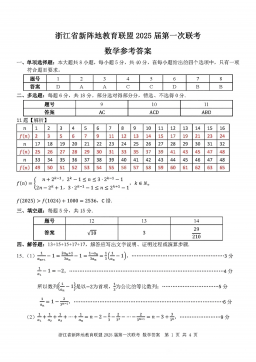
2025届浙江省新阵地联盟高三10月联考数学答案
分类:中学教育
时间:2025-12-31
标签:无
格式:PDF
价格:10 玖币
-
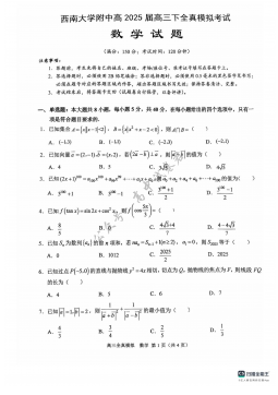
2025届重庆市西南大学附属中学高三下学期5月全镇模拟数学试题(含答案)
分类:中学教育
时间:2025-12-31
标签:无
格式:PDF
价格:10 玖币
-

2025届重庆康德三诊英语+答案
分类:中学教育
时间:2026-01-03
标签:无
格式:PDF
价格:10 玖币