Quantitative elemental imaging in eukaryotic algae Stefan Schmollinger12 Si Chen3 and Sabeeha S. Merchant12 1California Institute for Quantitative Biosciences QB3 University of California Berkeley CA
2025-05-02
12
0
1.39MB
33 页
10玖币
侵权投诉

Quantitative elemental imaging in eukaryotic algae
Stefan Schmollinger1,2,*, Si Chen3 and Sabeeha S. Merchant1,2
1California Institute for Quantitative Biosciences (QB3), University of California, Berkeley, CA
94720
2Departments of Molecular and Cell Biology and Plant and Microbial Biology, Berkeley, CA 94720
33X-ray Science Division, Argonne National Laboratory, Lemont, IL 60439
* Present address: MSU-DOE Plant Research Laboratory, Michigan State University, East
Lansing, MI 48824
Corresponding author: Stefan Schmollinger
612 Wilson Road, MSU-DOE Plant Research Laboratory, Michigan State University, East
Lansing, MI 48824, USA, +1 (310) 779-0687, schmolli@msu.edu
Keywords: trace metal sequestration, heavy metal detoxification, iron, copper, manganese, XRF,
SXRF, Chlamydomonas
Stefan Schmollinger (https://orcid.org/0000-0002-7487-8014)
Si Chen (https://orcid.org/0000-0001-6619-2699)
Sabeeha S. Merchant (https://orcid.org/0000-0002-2594-509X)

2
Abstract
All organisms, fundamentally, are made from the same raw material, namely the elements of the
periodic table. Biochemical diversity is achieved with how these elements are utilized, for what
purpose and in which physical location. Determining elemental distributions, especially those of
trace elements that facilitate metabolism as cofactors in the active centers of essential enzymes,
can determine the state of metabolism, the nutritional status or the developmental stage of an
organism. Photosynthetic eukaryotes, especially algae, are excellent subjects for quantitative
analysis of elemental distribution. These microbes utilize unique metabolic pathways that require
various trace nutrients at their core to enable its operation. Photosynthetic microbes also have
important environmental roles as primary producers in habitats with limited nutrient supply or toxin
contaminations. Accordingly, photosynthetic eukaryotes are of great interest for biotechnological
exploitation, carbon sequestration and bioremediation, with many of the applications involving
various trace elements and consequently affecting their quota and intracellular distribution. A
number of diverse applications were developed for elemental imaging allowing subcellular
resolution, with X-ray fluorescence microscopy (XFM) being at the forefront, enabling quantitative
descriptions of intact cells in a non-destructive method. This Tutorial Review summarizes the
workflow of a quantitative, single-cell elemental distribution analysis of a eukaryotic alga using
XFM.

3
The aim of this review is to (1) highlight the contributions of different elements to photosynthetic
life and the concepts of how organisms control their elemental composition, (2) introduce the
methodologies involved in studying elemental distributions in cells, especially X-ray fluorescence
microscopy (XFM, XRF), (3) review the current state of XFM studies in eukaryotic algae, and (4)
to extract a methodology frame-work for conducting XFM studies from these works. It is our goal
to facilitate the entry into the field of elemental research for algae scholars encountering questions
of metal homeostasis and elemental heterogeneity for the first time, and to encourage the use of
quantitative elemental imaging approaches for the purpose of determining biological function.
Elemental composition of cells
The elements of the periodic table are the indivisible foundation of all matter, including all
biological life of our planet (Figure 1). Every component of a cell is assembled from a selection of
elements, most prominently C, H, N, O, P, S, which are essential and constitute the backbone of
proteins, carbohydrates, nucleic acids and lipids (1-3). Essentiality of an element is defined by its
irreplaceability and its requirement in metabolism to complete the vegetative or reproductive life
cycle (4), while beneficial elements only improve the organisms fitness. The set of essential
elements therefore can vary between different organisms, depending on the organism’s
environmental niche and its required enzyme portfolio. It is estimated that ~ 40 % of all enzymes
utilize a uniquely suited element outside the group of macronutrients (CHNOPS) within their
catalytic centers to enabling catalysis (5, 6). Most organisms employ several cations (K, Mg, Ca
and to a lesser degree Na) and the Cl anion, all of which are abundant constituents of biological
matter, to regulate osmotic pressure and pH, build gradients across membranes that facilitate
energy production, transport processes or signal transduction, or serve as cofactors or allosteric
regulators in metabolites or proteins (7). And lastly, organisms require various additional sets of
elements in trace amounts (including many metal ions), to enable chemical functionalities that are
not provided from metabolites or amino acids (3, 8). These micronutrients/trace elements are

4
required co-factors for metabolically-essential enzymes, or for enzymes that enable new
biochemical capabilities, and consequently their acquisition, intracellular distribution and
utilization are a crucial aspect of cellular metabolism (3). Trace elements typically present the
most interesting targets for elemental imaging, because of the impact of their chemistry on cell
health and metabolism, the range of their abundances in an organism and the dynamic regulation
involved in their utilization (3, 9). A set of trace elements is essential for most organisms, including
Fe, Cu, Co, Ni, Mn, Zn, Se, V, I and Mo. Redox biochemistry is a central aspect of trace metal
utilization. Mn, Cu, and Fe, are therefore among the most abundant and important trace elements
for all organisms; at lower abundance Ni, Co and Mo are also required as cofactors by many
organisms (5, 10-17). Fe, Cu and Mn, among many important contributions, are critical for
photosynthetic electron transfer (18). Ni is used in a wide range of organisms, for example in
ureases and hydrogenases (19). Mo, outside of bacterial Mo-nitrogenase, is usually found in the
Molybdenum cofactor Moco, which is used in many enzymes, most prominently nitrate reductase
(17). Zn is similarly abundant and widespread as a trace element as Fe, Cu and Mn, but is used
as a Lewis acid and a structural component for proteins in most organisms (20-22). Se, most
prominently, is utilized as selenocysteine in specific enzymes requiring the element in their
catalytic centers, for example glutathione peroxidases (23-26). Co is used as a cofactor in a few
enzymes directly, but most famously is at the center of cobalamin (also known as vitamin B12),
which is critical to nitrogen-fixing bacteria (27, 28). V is used in vanadium-dependent
nitrogenases and haloperoxidases, and has an important role as an electron acceptor in
bacterial respiration (29). I is used in thyroid hormones in vertebrates and haloperoxidases in
algae (30, 31).
Othertrace elements (B, Si, As, Br, Sr, Cd, Ba, W, Hg, Pb, La, Ce and Nd) are either employed
in very specialized roles in select organisms in a specific environmental niche, or in individual
enzymes with a specific beneficial, but non-essential function (32-35).
5
Many of the trace elements, among other bioactive elements, can also accumulate in organisms
involuntarily, using uptake routes for intended cofactors, and interfere with biological processes
either in an advantageous, or more commonly a detrimental way.
Therefore, utilization of, especially the redox-active trace elements, comes with inherent risks.
The chemical reactivities that make these elements useful in the first place must be controlled
intracellularly to avoid unintended reactions (36-38). The concentration of many of these elements
in the environment of cells is a critical parameter, determining if the organism is starving for the
element as a nutrient, if either abundance or bioavailability is low, or if cell health is threatened by
overexposure (39). The toxicity can either be directly attributed to the detrimental reactivities of
the elements when uncontrolled, to the production of secondary toxic products, for example
reactive oxygen species, or via enzyme mis-metalation. Mis-metalation is largely attributed to the
inherent flexibility in proteins and the similar physical properties (ionic radii, charge and
coordination preferences) of the biologically common trace metal (40, 41). Most enzymes are
tuned to function with a specific metal cofactor. Binding of a different, similar metal at the active
site can result in loss-of-function, or worse, the production of unintended products or promotion
of side-reactions (38, 42, 43). All organisms therefore carefully control their elemental composition
at the point of uptake, resulting in specific cellular quotas, especially in the case of redox active
trace elements. Cells also employ elaborate strategies to avoid mis-metalation intracellularly,
including the compartmentalization of specific elements to ensure that the correct metal binds to
newly-synthesized proteins, or the use of metallochaperones to ensure correct delivery through
protein-protein interactions (44-46). Some metals are associated with organic groups or build into
large clusters (for example Fe in heme and Fe-S clusters) for similar reasons.
The pathways for trace element metabolism are among the most ancient in biology (47), and the
general concepts involved in trace metal utilization are well conserved across organisms.
Photosynthetic organisms specifically have unique requirements with respect to the elemental
composition, because of the metabolic demand of the photosynthetic apparatus and specific

摘要:
展开>>
收起<<
QuantitativeelementalimagingineukaryoticalgaeStefanSchmollinger1,2,*,SiChen3andSabeehaS.Merchant1,21CaliforniaInstituteforQuantitativeBiosciences(QB3),UniversityofCalifornia,Berkeley,CA947202DepartmentsofMolecularandCellBiologyandPlantandMicrobialBiology,Berkeley,CA9472033X-rayScienceDivision,Argonn...
声明:本站为文档C2C交易模式,即用户上传的文档直接被用户下载,本站只是中间服务平台,本站所有文档下载所得的收益归上传人(含作者)所有。玖贝云文库仅提供信息存储空间,仅对用户上传内容的表现方式做保护处理,对上载内容本身不做任何修改或编辑。若文档所含内容侵犯了您的版权或隐私,请立即通知玖贝云文库,我们立即给予删除!
相关推荐
-
机器人技术讲稿第八章 机器人编程VIP免费

 2024-12-10 13
 -
1,PCB专业用语VIP免费

 2024-12-10 13
 -
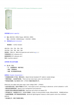
7,通信高频开关电源系统VIP免费
 2024-12-10 13
 -
单片微型计算机原理及接口技术第00章 目录VIP免费
 2024-12-10 16
 -
交大电气-信号与系统分析《信号与系统》期末-简单卷2000年VIP免费
 2024-12-10 15
 -
交大电气-信号与系统分析《信号与系统》期末-简单卷2001年VIP免费
 2024-12-10 15
 -
交大电气-信号与系统分析《信号与系统》期末-简单卷2002年VIP免费
 2024-12-10 14
 -
文献检索、调研、获取、管理方法医学文献和文献检索概论VIP免费
 2024-12-10 18
 -
文献检索、调研、获取、管理方法第九章__综合VIP免费
 2024-12-10 17
 -
传感器原理及工程应用第10章 超声波传感器VIP免费
 2024-12-10 16
分类:图书资源
价格:10玖币
属性:33 页
大小:1.39MB
格式:PDF
时间:2025-05-02
作者详情
相关内容
-

5.江苏省扬州市扬州中学高三年级物理学科高考模拟试卷
分类:高等教育
时间:2025-01-03
标签:无
格式:DOC
价格:5.9 玖币
-
4.湖南省衡阳市高三年级物理学科高考模拟试卷
分类:高等教育
时间:2025-01-03
标签:无
格式:DOC
价格:5.9 玖币
-
3.河北省沧州市第一中学高三年级物理学科高考模拟试卷
分类:高等教育
时间:2025-01-03
标签:无
格式:DOC
价格:5.9 玖币
-
2.广东省潮州市高三年级物理学科高考模拟试卷
分类:高等教育
时间:2025-01-03
标签:无
格式:DOC
价格:5.9 玖币
-
1.北京市第二中学高三年级物理学科高考模拟试卷
分类:高等教育
时间:2025-01-03
标签:无
格式:DOC
价格:5.9 玖币